Table of Contents
Dystrophy Fuchs – Explaining
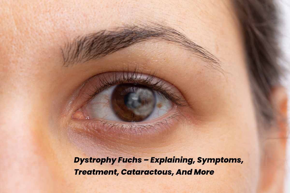
Dystrophy Fuchs is a corneal condition caused by a genetic mutation. Even though a patient is born with the disease, it is not detectable or symptomatic until later in life. The layer of cells responsible for maintaining adequate fluid levels in the cornea (endothelium) will degrade as the condition progresses.
Causing small bumps (guttae) to grow on the rear of the cornea. Fluid builds up in the cornea when enough cells are lost, producing oedemic. Corneal oedemic, or swelling of the cornea, causes eyesight clouding or blurring.
Symptoms of Dystrophy Fuchs
Blurred vision is one of the initial indicators of Fuchs’ dystrophy, caused by fluid accumulation in the cornea. Excess fluid builds up overnight during sleep in the early stages of the condition, producing hazy vision and pain when you wake up. Over the day, extra juice can be squeezed out of the cornea, lowering swelling and increasing vision. This may go on for hours.
Swelling, poor vision and pain remain longer in severe stages of Fuchs’, often up to daily.
Other signs and symptoms include:
Rough or gritty sensations in the eyes, sometimes accompanied by severe pains in the eyes
- Discomfort when exposed to bright light
- Eyesight that fluctuates throughout the day or from day to day, generally worse in the morning or on humid, rainy days
- Bright lights can cause halo effects and glares.
- The Color contrast is low due to blurry vision.
Fuchs’ Dystrophy Diagnosis
During a routine eye checkup, Fuchs’ dystrophy is frequently detected. Your eye doctor may perform the tests listed below to confirm a diagnosis.
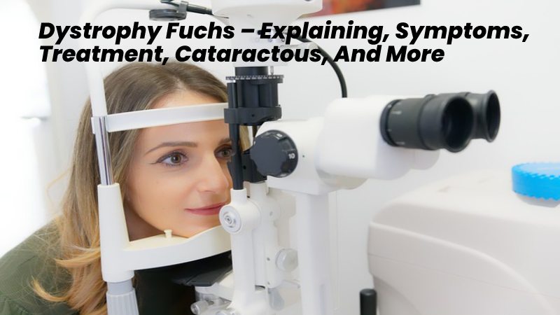
Slit-lamp microscopy: A slit lamp provides a light beam that may be changed from a tiny slit to a circular circle, depending on whatever portion of the eye the doctor wishes to examine. The connected microscope enables for high magnification viewing of eye anatomy.
The pachymeter, which measures corneal thickness, is essential in diagnosing illnesses that cause the cornea to thicken, such as Fuchs’ dystrophy.
- Confocal/specular microscopy: A confocal/specular microscope beams light onto the corneal endothelium, allowing for photography. It can count, count, count, count, count, count, count, count, count, count, count, count, count, count, count, count, count, count, count, count, count
Fuchs’ Dystrophy Treatment
While there are currently no treatments that can reverse Fuchs’ dystrophy, non-surgical treatments such as eye drops or eye ointment can help in the condition’s early stages.
More Advanced Treatment
If the condition progresses and interferes with daily tasks, your doctor may recommend it. Eye drops or ointments that draw fluid out of the cornea are used to relieve symptoms of Fuchs dystrophy. If painful sores develop on the cornea, soft interaction lenses or surgery to create flaps done the sores may help reduce pain.
The only cure for Fuchs Dystrophy is a corneal Transplant.
Until recently, the most common type of corneal transplant was powerful keratoplasty. The procedure, a small round piece of the cornea, leaves an opening in the front of the eye. A part of the cornea from a human donor is then sewn, opening in front of the eye.
A newer method called endothelial keratoplasty (DSEK, DSAEK, or DMEK) has developed the favourite option for people with Fuchs dystrophy. Only the cornea’s inner layers are replaced in this procedure instead of all the layers. This leads to a faster repossession and fewer complications. Stitches are most often not needed.
Surgical Procedures Such As:
- Corneal transplantation
- Endothelial keratoplasty is a procedure that removes the outer layer of the cornea. The corneal endothelium and Descemet’s membrane. Compromise by Fuchs’ dystrophy, are replaced with healthy corneal tissue from an organ donor during this corneal transplant surgery. Descemet stripping automatic endothelial keratoplasty (DSAEK) and Descemet’s casing endothelial keratoplasty are two prevalent types of endothelial keratoplasty (DMEK).
Cataractous and Fuchs’ Dystrophy
Cataracts can develop in people with Fuchs’ dystrophy. Cataract surgery may be the sole option in mild or moderate Fuchs’ severity with cataracts. Corneal transplantation requires after cataract surgery if:
- Following cataract surgery, recovery is sluggish or non-existent.
- Following surgery, Fuchs’ dystrophy condition worsens.
Cataract operation and a corneal transplant may indicate simultaneously in severe Fuchs’ dystrophy situations. Patients benefit from a faster overall healing time when using a combination strategy.
Based on a comprehensive examination, your ophthalmologist will propose a treatment strategy.
Surgery for Dystrophy and Vision Correction
Elective laser vision correction recommends for patients with dystrophy. The cornea’s compromised state might deteriorate, affecting the treatment’s result.
Causes
Fuchs dystrophy inherits which approve unhappy parents to children. If also of your parents has the disease, you have a 50% chance of developing the disorder.
However, the disorder may also occur in people deprived of a recognized domestic past of the disease.
Fuchs dystrophy is more common in women than in men. Vision problems do not appear before age 50 years in most cases. Though, a health care provider may see signs of the disease in affected people in their 30s or 40s.
Fuchs dystrophy affects the high layer of cells that appearances the back part of the cornea. These cells help pump extra fluid out of the cornea. As extra and extra cells are lost, the liquid starts to shape up in the cornea, causing a bulge and a cloudy cornea
At first, liquid may build up only during sleep, once the eye. As the disease becomes worse, small swellings may form. The blisters get more extensive and may ultimately break. This causes eye pain. can also cause the formation of the cornea to alteration, leading to more dream difficulties.
Conclusion
Fuchs dystrophy gets worse over time. Without a corneal transplant, a person with severe may become blind or have severe pain and significantly reduced vision.
Mild cases often worsen after cataract surgery. A cataract surgeon will evaluate this risk and may modify the technique or the timing of your cataract surgery.