Ultrasound An ultrasound is an imaging test that uses sound whitecaps to create pictures of structures, tissues, and other constructions inside the body. An ultrasound can also show body helpings in motion, such as a heart beating or blood flowing through blood vessels. Unlike X-rays, ultrasounds don’t use any radiation.
There are two main groups of ultrasounds: pregnancy ultrasound and methodical ultrasound.
A pregnancy ultrasound is used to look at an unborn baby. The test can offer information about a baby’s growth, development, and general health.
Diagnostic ultrasound is used to view and offer info about other inside parts of the body. These contain the heart, blood vessels, liver, bladder, kidneys, and female generative structures.
Table of Contents
Ultrasound Uses
An ultrasound can be used in others, depending on the type of ultrasound and which part of the body is being checked.
A pregnancy ultrasound is done to get material about the well-being of an unborn baby. It may be used
Confirm that you are expectant.
Check the size and location of the unborn baby.
I squared to see you are pregnant with more than one baby.
Approximation of how long you have been expecting. This is known as gestational age.
Check for signs of Unhappy condition, which include thickening in the back of the baby’s neck.
Check for congenital disabilities in the brain, spinal cord, heart, or other parts of the body.
Amniotic fluid is a clear melt that surrounds an unborn baby during pregnancy. It defends the baby from outside injury and cold. It also helps encourage lung development and bone growth.
Diagnostic Ultrasound May be Used to
Invention out if blood is flowing at a normal amount and level.
See if there is a problem with the construction of your heart.
Look for blockages in the gallbladder.
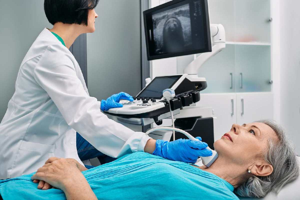
I squared the thyroid gland for cancer or non-cancerous evolutions.
Check for irregularities in the stomach and kidneys.
Help guide an operation process. An operation is a procedure that removes a small example of tissue for testing.
In women, problem-solving ultrasound may be used to:
Look at a chest lump to see if it strength be cancer.
In men, diagnostic ultrasound may help diagnose disorders of the prostate gland.
Needs an Ultrasound
You may need a diagnostic ultrasound if you have symptoms in specific organs or tissues. These contain the heart, kidneys, thyroid, gallbladder, and female generative system. You may also need an ultrasound if you are getting a biopsy. It helps your health care worker get a clear image of the area that test.
The provisions will rest on which type of ultrasound you are having. For ultrasounds of the abdominal area, including condition ultrasounds and ultrasounds of the female generative classification, you may require to fill up your bladder before the test.
Some types of ultrasounds need no preparation at all. This involves drinking two to three water spectacles about an hour before the test, and not going to the bathroom. For other ultrasounds, you might need to adjust your diet or fast for several hours before your examination.
Your well-being care worker will let you know if you need to do anything to prepare for your ultrasound.
Ultrasound Scans Work
Ultrasound scans, or sonography, are safe since they use sound waves or echoes to make an image instead of radiation. An ultrasound scan uses high-frequency sound whitecaps to take pictures of the inside of the body. It is suitable for use during pregnancy.
The scans use to evaluate fetal growth, and they can detect difficulties in the liver, heart, kidney, or abdomen. They may also assist in the performance of certain types of biopsies.
Fast Facts on Scans
Pregnancy. Ultrasound images have many uses through pregnancy. Early on, they might use to determine due dates. Expose the attendance of twins or other multiples, and rule out ectopic pregnancies. They also are valuable broadcast tools in helping to detect possible problems, including some congenital disabilities, placental issues, breech placing, and others.
Many expectant parents look onward to learning the sex of their babies halfway done a pregnancy. And later in pregnancy, doctors can smoothly use to evaluate how large a baby is previously distribute
Diagnostics. Doctors employ imaging in identifying a complete change of situations moving the structures and soft matters of the body, including the heart and blood bowels, liver, gallbladder, spleen, pancreas, kidneys, bladder, uterus, fruits, eyes, thyroid, and testicles. have some diagnostic limits. However, sound whitecaps do not transmit well through dense bone or body parts that may hold air or gas, such as the bowel.
Ultrasound scans are safe and extensively used.
They use to check the progress of a pregnancy.
They use for diagnosis or treatment.
No special preparation is ordinarily necessary before a scan.
Ultrasound Transducers
The transducer or on the surface of the patient’s body, but some kids place it internally.
Examples are
an endovaginal transducer for use in the vagina
an endorectal transducer for use in the rectum
a transoesophageal transducer passed down the patient’s throat for use in the oesophagus
Some tiny transducers place onto the end of a tube and introduced into blood vessels to inspect the walls of blood vessels.
Types
Most ultrasounds use a transducer on the external of the skin. Occasionally, however, doctors and technicians can get a better diagnostic image by introducing a special transducer into one of the body’s natural openings:
A transducer stack in a woman’s vagina to improve images of their uterus and fruits in a transvaginal. A transrectal is occasionally in the diagnosis of prostate conditions.
A transoesophageal echocardiogram uses the transducer probe in the exophages so that the sonographer can obtain more explicit images of the heart. Moreover, technology has progressed to allow for different types of imaging:
Bone sonography helps doctors identify osteoporosis. Echocardiograms to view the heart. Doppler is a particular type that creates images of blood flow over vessels.
3D imaging adds extra length to the image, creating three-dimensional clarifications rather than the flat images made with traditional.
4D ultrasounds show 3D images in motion.
Conclusion
Studies have shown that it is usually safe. There are no known damaging side effects, and there is virtually no discomfort during the test. In accumulation, does not use energy, as X-ray tests do. The possible A typical takes between 30 minutes and an hour. Ultrasounds are usually uncomfortable, and you are awake and alert during the procedure. Often a technician will discuss what they see during the test, but in some instances, you may need to wait to discuss the conclusions with your doctor.
Relate Searches:
ultrasound scan
types of ultrasound
ultrasound machine
ultrasound pregnancy