Computed Tomography scan ( CT scan) of the cervical spine is a diagnostic test that uses X-rays to produce detailed images of the cervical spine, which includes the seven vertebrae in the neck. CT scans can provide detailed information about the bones and soft tissues in the cervical spine, including the discs, ligaments, and nerves.
The test is typically used to diagnose injuries, tumors, or other conditions that affect the cervical spine, such as degenerative disc disease or herniated discs. It is a non-invasive procedure typically performed in a hospital or imaging center. The results are usually interpreted by a radiologist or other medical professional with specialized training in interpreting imaging tests. Look for ct scan center near me to diagnose an injury or any other condition related to the cervical spine.
Table of Contents
What Is Spinal CT Scanning?
Computed tomography, sometimes a CT or CAT scan, is a diagnostic medical imaging procedure. It provides several pictures of the inside of the body, similar to standard x-rays.
Images from the spinal CT scan can be reformatted in multiple planes. It is even capable of producing three-dimensional visuals. These images can be reviewed on a computer display, printed on film or using a 3D printer
Spinal CT scans of internal organs, soft tissue, bone, and blood arteries show more information than standard x-rays. The bone structure of the spine, vertebrae, intervertebral discs, and, to a lesser extent, spinal cord soft tissues are clearly and precisely displayed with CT.
Purpose Of Spinal CT Scan
The CT scan can be used to diagnose a wide range of situations that affect the cervical spine, including:
- Herniated discs
- Bone spurs
- Spinal stenosis
- Fractures
- Tumors
- Infections
One of the significant advantages of a CT scan is its ability to provide detailed images of the cervical spine, which allows for accurate diagnosis of conditions that affect the bones and soft tissues. CT scans can also be used to plan surgery, such as spinal fusion or removing tumors or bone spurs.
The CT scan can also be used to monitor the progression of a condition over time. For example, if a patient has a herniated disc, a CT scan can be used to track the size of the herniation and monitor any changes in the size over time.
Preparation For The Spinal CT Scan
Wear loose-fitting, comfortable attire for your exam as you may need to shift into a gown for this CT scan procedure.
Metal objects, such as jewelry, dentures, eyeglasses, and hairpins, might interfere with CT pictures. So, remove them before your exam. Some CT scans will necessitate the removal of hearing aids and detachable dental work. Women will be required to remove bras with metal underwires and any piercings, if possible.
If your exam utilizes contrast material, your doctor may urge you not to drink or eat anything for a few hours before the exam. In addition, inform your doctor about all medications you are taking and any sensitivities you may have.
If you have any allergies to contrast material, your doctor may prescribe drugs (typically a steroid) to lessen your chances of having an allergic reaction. Contact your doctor before your exam appointment to avoid unnecessary delays.
Inform your doctor of any recent diseases and infections or other medical issues, as well as any family history of heart disease, asthma, diabetes, kidney disease, or thyroid problems. Any of these situations may enhance the likelihood of a negative impact.
Women should always notify their doctor and the CT technician if they suspect they are pregnant. Connect to the diagnostic test center near me for appropriate and timely report.
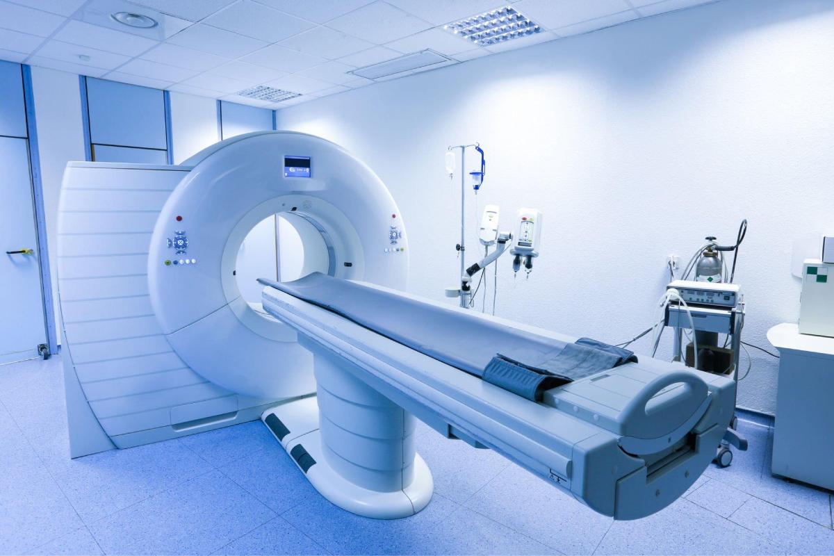
Procedure Of The Spinal CT Scan
The procedure typically takes about 15-30 minutes. First, the patient will be asked to change into a hospital gown and remove any metallic objects or jewellery that might interfere with the scan.
The patient will be positioned on a table that moves through a ring-shaped CT scanner during the CT scan. The scanner takes multiple X-ray images of the cervical spine as the table moves through the scanner. The patient may be asked to hold their breath for a few moments during the scan to ensure that the images are clear and sharp. In addition, the CT scanner is equipped with a device that will help the patient stay still during the scan.
Let the technologist know if the patient has any allergies or has ever reacted to the contrast material used for X-ray procedures. The scan may require an iodine-based contrast material to enhance the visibility of specific structures. The contrast material is usually given through an IV line placed in the patient’s arm.
After the CT scan is completed, the patient can return to normal activities. A radiologist will analyze the images and then send a report to the referring physician. The radiologist will look for signs of injuries, tumors, or other conditions affecting the cervical spine. The patient’s physician will review the results, and the patient will be informed of the developments.
Risk And Complication Of The Spinal CT Scan
CT scans of the cervical spine are generally considered safe, with minimal risk of complications. However, as with any diagnostic test that uses ionizing radiation, there is a small risk from radiation exposure. The risk from a CT scan is low, but it is still essential to consider the consequences and benefits of the test before undergoing the procedure.
It is also important to note that CT scans of the cervical spine are not recommended for pregnant women, as radiation exposure may harm the developing fetus.
Final Words
The Spinal CT scan is a diagnostic test that uses X-rays to produce detailed images of the cervical spine, which includes the seven vertebrae in the neck. CT scans can provide detailed information about the bones. The soft tissues in the cervical spine, including the discs, ligaments, and nerves. CT scans can diagnose a wide range of conditions that affect the cervical spine, including herniated discs, bone spurs, spinal stenosis, fractures, tumors, and infections. Moreover, CT scans can also be used to monitor the progression of a condition over time.
The CT scan is generally considered safe, with minimal risk of complications. However, as with any diagnostic test that uses ionizing radiation, there is a small risk of cancer from radiation exposure. Look for ct scan center near me to diagnose an injury or any other condition related to the cervical spine.